


Geological observations and laboratory analyses are an inseparable part of the Misliya Cave excavation project, in accordance with prehistoric research strategies firmly established in Israel since the 1990s. The integration of geology and archaeology, named geo-archaeology, provides deeper insights regarding the reconstruction of past human behavior, interactions between nature and culture, and site-formation processes.
During the period of human occupation, Misliya Cave was permanently filled with human-related debris and waste materials mixed with various natural deposits, which accumulated through wind action, slope processes, and/or karstic processes. Over time, remains of past human activities, e.g. hearths, may be eroded and vanish from the record. In other cases, luckily for geo-archaeologists, they may become incorporated and preserved in cave deposits. However, preservation of pristine archaeological features in cave deposits is always partial. In Misliya Cave these features are sometimes particularly hard to recognize in the indurated breccia-like layers because of strong secondary cementation, the result of precipitation from karstic, carbonate-bearing solutions that abound during intensive rains.
One of the most effective techniques to unravel the geological and human habitation history from the depositional record is micromorphology. Micromorphology is a study of tiny, microscopic features, which escape identification with the naked eye in the field and require the employment of additional laboratory techniques. Thus, geo-archaeologists take undisturbed geological samples from stratigraphical sections or specific site features in the dig for detailed microscopic analysis. Thin sections are prepared after impregnation of the undisturbed samples with special resins, then sliced and polished to 30 µm thickness. At such a thickness minerals, including colored ones, become transparent for transmission light microscopy. Usually we use magnification from 20x through 400x to precisely identify the nature of particular elements. Diagnostic micromorphological features are carefully documented and photographed. Examples of these are given below. Polished thin sections may be further analyzed by scanning electron microscope (SEM) equipped with a microprobe analyzer so as to enable the identification of the chemical composition of any feature that a geo-archaeologist is interested in.
In these ways we were able to identify a suite of features in the Misliya brecciated deposits that shed light on how the cave was used and what natural processes influenced the preservation of pristine features.
The Misliya Cave deposits are rich in animal bones, studied by zooarchaeologists, which detail the hunting repertoire of ancient humans. Likewise, remnants of fragmented and weathered chips of bones, ranging from 50µm to several mm thick are ubiquitous in thin sections. Micromorphologically, chips of bones show a broad range of coloration from pale-yellow to dark brown (Figure 1), probably due to heating. Bright brown tiny fragments of bones are mostly common (Figure 2).
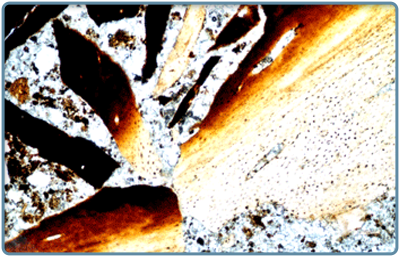 Figure 1
Fragmentation of bones is common for breccias. Bones also show weathering and probably heating effects as seen in colors changing from pale-yellow to dark brown
(magnification X40).
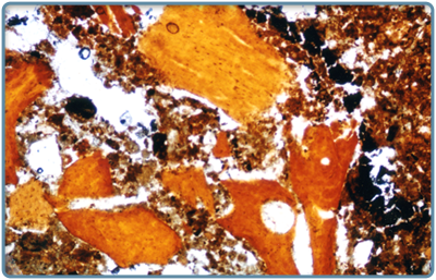 Figure 2
Brown colored fragmented chips of bones embedded in the clay soil mass with scattered charred materials
(magnification X40).
In addition to burnt bone fragments, thin sections allow us to identify a variety of combustible materials such as wood and grass. Figure 3 shows diverse charred materials appearing in black. Significantly, an upper black feature under twice as high magnification as in Figure 3 (Figure 4) clearly shows the cellular structure, which is an unambiguous indication of vegetal tissues.
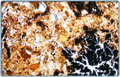 Figure 3
Charred materials embedded in the heterogeneous fine-grained mass of the breccia. The upper charred piece is shown in more detail in Figure 4
(magnification X40).
 Figure 4
Close-up view of a charred vegetal matter with preserved cellular structure
(magnification X100).
Thin sections exhibit an abundance and variety of calcite precipitations both in the matrix and in pore spaces. These observations are in good agreement with our earlier studies of lithified deposits from the cave (Weinstein-Evron et al., 2003). We found evidence of post-depositional travertine formation, which is represented by euhedral sparitic crystals, showing a quasi-radial pattern (Figures 5, 6).
 Figure 5
Secondary deposits of regular large calcite crystals, accumulating in a pore in a rosette-like form
(magnification X40).
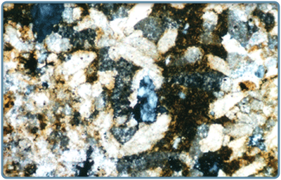 Figure 6
The same view as in Figure 5 under crossed polarized light, which allows us to better see the area of the breccia where large calcite crystals precipitated during karstic processes
(magnification X40).